Klassifikation Mammakarzinom
22.07.2021
Klassifikation Mammakarzinom
B-Klassifikation
| B-Kategorie | Beschreibung |
|---|---|
| B 1 | Nicht verwertbar oder ausschließlich Normalgewebe |
| B 2 | Benigne, u.a. fibrös-zystische mastopathie, Fibroadenom, sklerosierende Adenose, periduktale Mastitis |
| B 3 | Benigne, aber mit unsicherem biologischen Potential u.a. atypische intraduktale Epithelproliferationen bei denen eine definitive Festlegung an der perkutanen Biopsie nicht möglich ist (z. B. atypische duktale Hyperplasie: in Abhängigkeit von Ausdehnung und Grad der Atypie ggf. auch Kategorie B4); atypische lobuläre Hyperplasie und LCIS; papilläre Läsionen (bei hochgradigem V.a. papilläres DCIS: gegebenenfalls auch Kategorie B4); radiäre Narbe/komplexe sklerosierende Läsion; V.a. Phylloides-Tumor |
| B 4 | Malignitätsverdächtig z. B. vermutlich maligne Veränderung, aber Beurteilung aus technischen Gründen eingeschränkt; atypische intraduktale Epithelproliferationen in Abhängigkeit von Ausdehnung und Schwere der Atypie (siehe auch Kategorie B3) |
| B 5 | Maligne z. B. DCIS, invasive Karzinome, maligne Lymphome B5a= nicht-invasives Mamma-CA B5b= invasives Mamma-CA B5c= fraglich invasives CA B5d= maligner Tumor, nicht primär Mamma |
Dieses Beurteilungsschema, dass von der E.C. Working Group on breast screening pathology und der National Coordinating Group for Breast Screening Pathology (NHSBSP), Gro0ßbritannien, empfohlen wird, ist an zytologische Klassifikationsschemata angelehnt. Im Hinblick auf eine ausführliche Erläuterung der Bewertungskriterien wird auf die „Guideslines for non-operative diagnostic procedures and reporting in
breast cancer″ der NHSBSP verwiesen.
NHS Breast Screening Programme
Van Nuys - Score
| Index für therapeutische Empfehlungen des DCIS | |||
|---|---|---|---|
| Score | 1 | 2 | 3 |
| [1] Größe in mm | £ 15 | 16-40 | ³ 41 |
| [2] Breite der tumorfreien Ränder | ³ 10 | 1-9 | <1 |
| [3] Klassifikation | non-high-grade ohne Nekrosen | non-high-grade mit Nekrosen | high-grade mit Nekrosen |
| Score=[1]+[2]+[3] | Behandlung |
|---|---|
| 3,4 | therapiert durch segmentale Excision |
| 5,6,7 | segmentale Exzision + Strahlentherapie, ggf. Nachexzision |
| 8,9 | Mastektomie |
Score nach Silverstein et al. 1996 [6]
IRS
Teroidrezeptorstatus und Immunreaktiver Score
Semiquantitative Methode nach Remmele u. Stegner (vgl. auch Remmele Bd. 4, S.259)
| Färbeintensität (staining intensity = SI) | Score |
|---|---|
| keine Färbung nachweisbar | 0 |
| geringe Färbeintensität der Kerne | 1 |
| mäßige Färbeintensität der Kerne | 2 |
| starke Färbeintensität der Kerne | 3 |
| Anzahl positiver Zellen (PP) | Score |
|---|---|
| keine positien Kerne nachweisbar | 0 |
| weniger als 10% positiver Zellen (Kernfärbung) | 1 |
| 10-50% positive Zellen (Kernfärbung) | 2 |
| 51-80% posiive Zellen (Kernfärbung) | 3 |
| mehr als 80% positiver Zellen (Kernfärbung) | 4 |
| Bewertung des immunreaktiven Scores (IRS=SI * PP) |
Score |
|---|---|
| negativ | 0 |
| 1 | |
| 2 | |
| positiv | 3 |
| 4 | |
| 6 | |
| 8 | |
| 9 | |
| 12 |
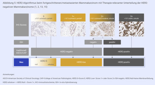
Her-2/neu
Interpretation der Her-2/neu-Immunhistochemie
| Staining Pattern | Score | HER2 Overexpression Assessment (Report to treating physician) |
|---|---|---|
| No staining at all, or membrane staining in less than 10% of the tumour cells is observed. |
0 | Negative |
| A faint/barely perceptible membrane staining is detected in more than 10% of the tumour cells. The cells are only stained in part of their membra |
1+ | Negative |
| A weak to moderate staining of the entire membrane is observed in more than 10% of the tumour cells. |
2+ | Weakly positive |
| A strong staining of the entire membrane is observed in more than 10% of the tumour cells. |
3+ | Strongly positive |
| score: 0 - negativ | score: 0 - negativ | |
 |
 |
|
| score 1 + - negativ | score 1 + - negativ | |
 |
 |
|
| score 2 + - schwach positiv | score 2 + - schwach positiv | score 2 + - schwach positiv |
 |
 |
 |
| scrore 3 + - stark positiv | scrore 3 + - stark positiv | scrore 3 + - stark positiv |
 |
 |
 |
Photos by James Thompson, MD, PhD, Director of Pathology, Biopharmaceutical Services, Impath Lab., Frolian Espinoza, MD, Molecular Tissue Pathology,
Quest Diagnostics/Nichols Institute and DAKO
Wann ist eine HER-2/neu-FISH-Analyse nötig?

 |
 |
| HER2 FISH - kein amplifiziertes HER2-Gen | HER2 FISH - amplifiziertes HER2-Gen |
CISH analog zu FISH ohne qualitativen Unterschied:

Interpretation:
Anzahl der Signale pro Kern: Mittelwert aus 2x20 ausgezählten Tumorzellkernen.
| Kriterium | Signale pro Kern |
|---|---|
| normal | < oder = 4 |
| low level - Amplifikation | 5-10 |
| high leveel - Amplifikation | mehr als 10 oder große Cluster |
Regressionsgrad n. Sinn
| Regressionsgrad | Morphologie |
|---|---|
| 0 | kein morphologischer Effekt |
| 1 | vermehrte Tumorsklerose mit herdförmiger resorptiver Entzündung und/oder deutlichem zytopathischen Effekt |
| 2 | weitgehende Tumorsklerose mit nur fokal noch nachweisbarem, evtl. auch multifokalem, minimal invasivem Resttumor (< = 0,5 cm ), häufig ausgedehnte intraduktale Tumorausbreitung |
| 3 | kein invasiver Resttumor |
| 4 | kein Resttumor |
